 Microscopic Tests Malaria Site from i2.wp.com
The malaria parasite is spread by female anopheles mosquitoes. This is an open access article distributed under the terms of the creative commons attribution. A drop of blood from the patient is spread on a slide and stained with giemsa stain. Thick blood film samples a relatively large volume of blood thus allowing more efficient detection of parasites (increased sensitivity). In all stages, however, the same you will need to refocus, using the fine adjustment, each time you move the microscope field: 6 867 просмотров 6,8 тыс. It causes malaria, which has been shown to present significant health risks to pregnant women and. Viewing a human parasite under the microscope is observing an organism that lives in (or on) another living organism (host) for survival. The direct microscopic visualization of the malarial parasite on the thick and/or thin blood smears has been the gold standard for malaria diagnosis. Human lab workers would mostly focus on preparing the slides of blood.
Stained blood filmsthe accepted laboratory practice for the diagnosis of malaria is the preparation the sensitivity of ao staining for detection of malaria parasites in infections with parasite levels of the manufacturing of rdt is currently under review by the larger companies (amrad ict and.
The parasite positivity of malaria by using partec rapid malaria test was 88 (48.9%). Physicians aren't aware of how widespread parasitic infections are. This is then stained and examined carefully under a microscope. Viewing a human parasite under the microscope is observing an organism that lives in (or on) another living organism (host) for survival.
 Source: www.researchgate.net
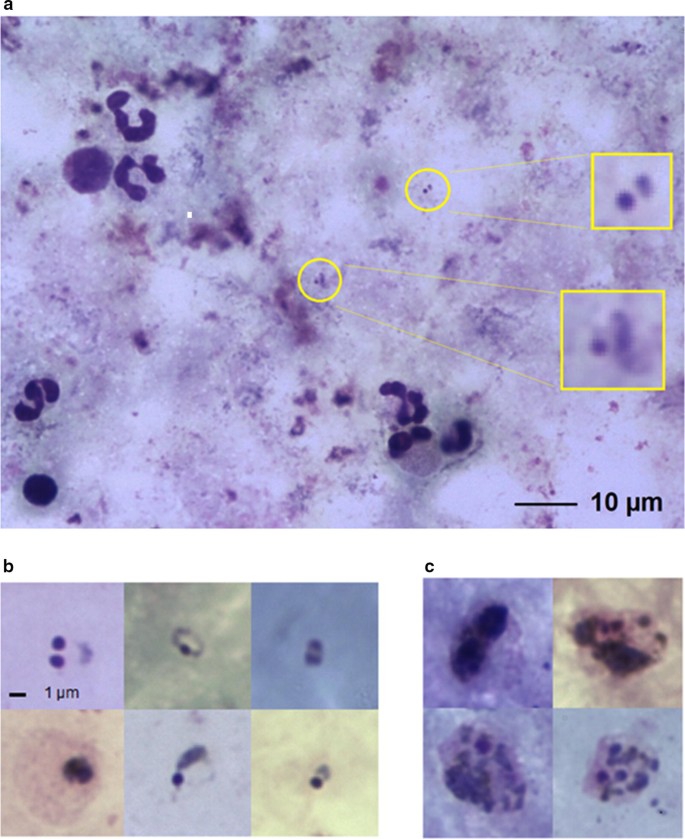
Leishman A And Giemsa B Stained Peripheral Blood Thick Smear Download Scientific Diagram
 Source: upload.wikimedia.org
Plasmodium Vivax Wikipedia
 Source: helid.digicollection.org
Basic Malaria Microscopy Part I And Ii Learning Unit 8 Examining Blood Films For Malaria Parasites
 Source: i.ytimg.com
Geimsa Stain For Plasmodium And Babesia Parasite Diagnostics Youtube
 Source: www.pathologyoutlines.com
Pathology Outlines Plasmodium Falciparum
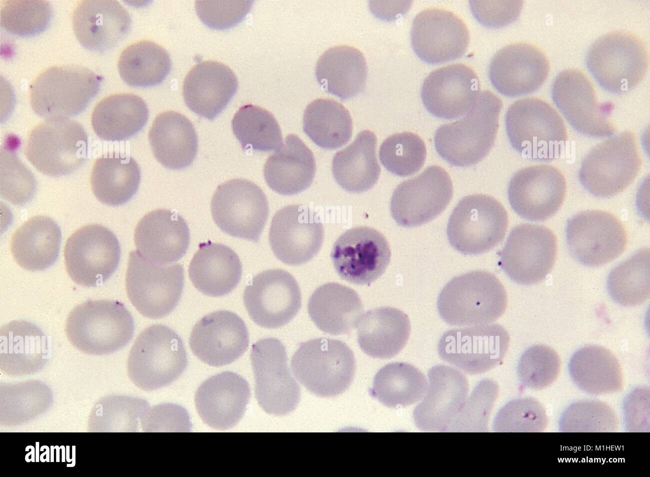 Source: c8.alamy.com
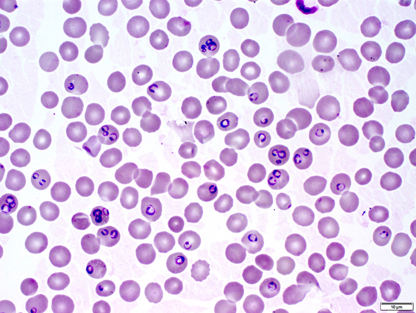
Malaria Parasite Revealed In A Debris Of Red Blood Cell Using Giemsa Stock Photo Alamy
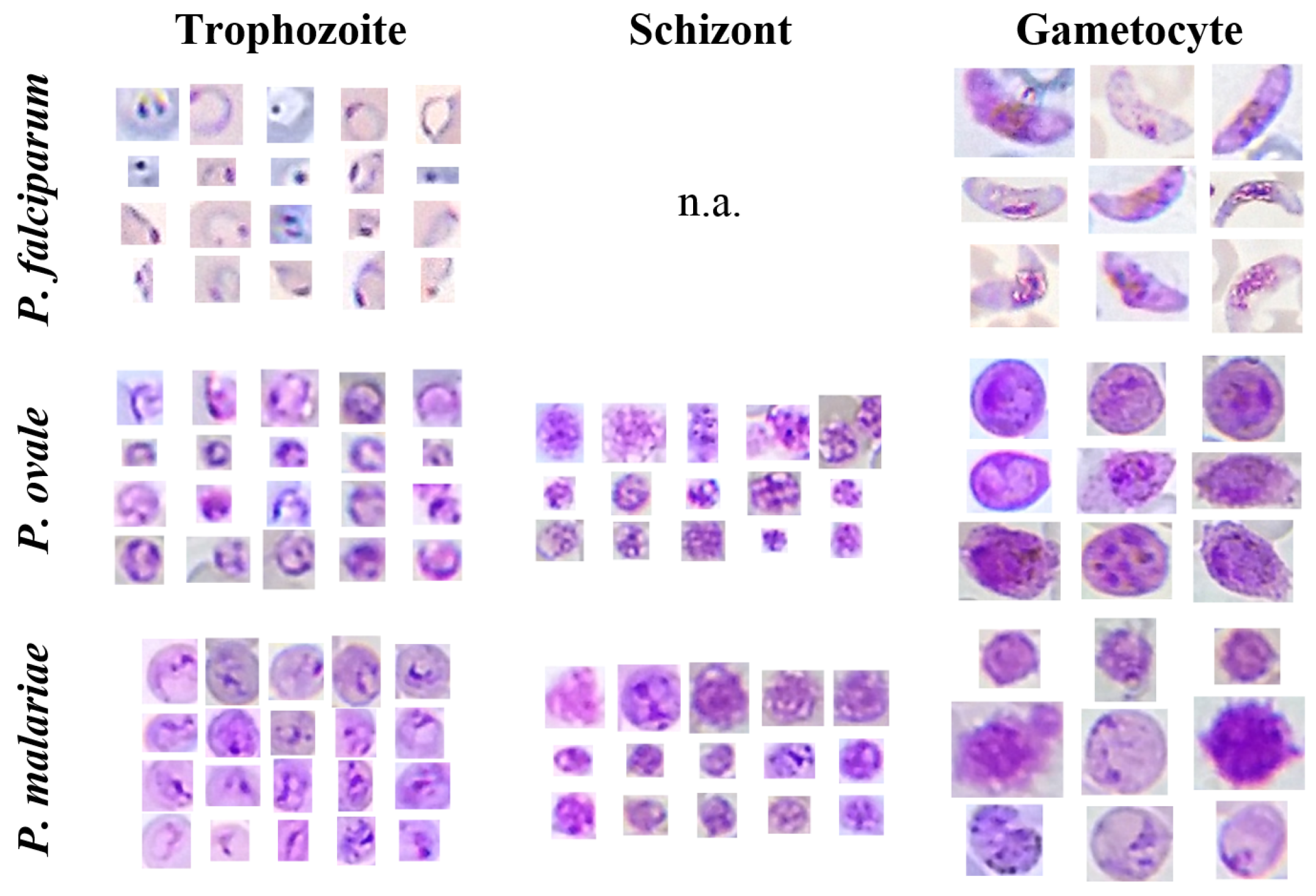 Source: www.mdpi.com
Sensors Free Full Text Mobile Based Analysis Of Malaria Infected Thin Blood Smears Automated Species And Life Cycle Stage Determination Html
 Source: img.bioopticsworld.com
Label Free 3d Microscopy Method Furthers Malaria Research Biooptics World
 Source: media.springernature.com
Automated Microscopy For Routine Malaria Diagnosis A Field Comparison On Giemsa Stained Blood Films In Peru Malaria Journal Full Text
For more than hundred years, the direct microscopic visualization of the parasite on the thick and/or thin blood smears has been the accepted method for the diagnosis of malaria in most. When viewed under blue light (~460 nm), parasites stained with acridine orange will fluoresce brightly the qbc malaria test is designed to work with a fluorescence microscope. The parasites are very small (microscopic) and can be seen only under a microscope with high when the microscopist sees stained parasites, the diagnosis of malaria is confirmed.
Get Latest Ideas : HOME